Idea for Efficient Cell Image Compression
Premise
Pathology and histology slide images are taken with extremely high resolution cameras, resulting in:
- High cost of storage
- Bandwidth issues while transporting images
It is important to note that these images often rely on lossless compression, because any artifacting will result in a lowered ability for the doctor to give accurate results.
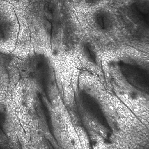
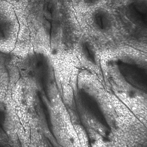
Example of a cell image.
Proposed Solution
In most regions of the body, neigboring cells looks alike to the cells bordering it. I propose the following solution to compress the images:
- Segment the image into distinct cells.
- Take a dot product of the matrix of cells with itself (ATA) to figure out which cells are most similar to other cells. I call these “reference cells”.
- Store a set of deltas for all other cells in terms of rotations, translation, and transformations.
- Apply further compression using any standard algorithm to the deltas themselves.
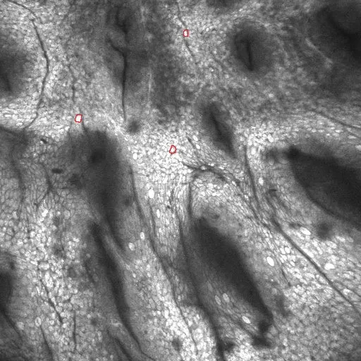
Example of "reference" cells.
A set of deltas in a binary format would be better than a large amount of pixels and allow for better compression.
I attempted to do something along these lines on GitHub here, but had to stop development due to time constraints. Hopefully I can continue this down the line!